

Clone ID / Name: 7G10 anti-Fasciclin III
Anti - Fasciclin III (Drosophila)
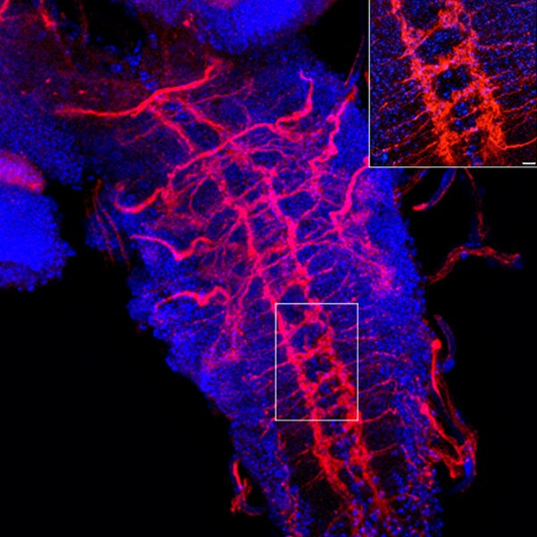
Confocal microscope image of a Drosophila larva ventral nerve cord labeled with DSHB antibody 7G10 anti-Fasciclin III (red). PMID: 31213988, Fig. 3B.
Catalog Fields
Identity
Clone ID / Name: 7G10 anti-Fasciclin III
Alternate Antibody Name: —
Antigen Name: Fasciclin III (Drosophila)
Alternate Antigen Name: —
Gene Symbol: —
Gene Name: Fas3
Entrez Gene ID: 35097
Antigen Sequence: —
Antigen Species: Drosophila
Antigen Mol. Weight: Multiple isoforms; Apparent: 46, 59, 66, and 80 kDa
Uniprot ID: P15278
Human Protein Atlas: —
Antibody Properties
Host Species: Mouse
Clonality: Monoclonal
Isotype: Mouse IgG2a
Myeloma Strain: NS-1
Antibody Registry / RRID: AB_528238
Available to For-Profits: Yes
Hybridoma Available: Yes
Cell lines cannot be distributed to commercial/for-profit users.
Cell lines cannot be distributed to commercial/for-profit users.
Immunogen & Epitope
Immunogen: Membrane preparation from nerve cord and cultures of primary neurons (10-13 hr development).
Immunogen Sequence: —
Epitope Mapped: No
Epitope Location / Seq: —
Clone: —
Additional Info: This antibody may work for Immunoprecipitation. While the Depositor clearly stated that it doesn't work for immunoprecipitation (see Depositor Notes), the Initial Publication, in which the Depositor is the last author, reports the succesful use of 7G10 for the immunoprecipitation of Fasciclin III.
Add. Characterization: —
Species Reactivity
Confirmed Reactivity: Drosophila
Predicted Reactivity: —
Depositor
Depositor Name: C. Goodman
Institution: University of California, Berkeley
Deposit Date: February 1, 1994
Depositor Notes: Fas III is expressed on a subset of neurons and axons in the CNS, patches of ectoderm, ectodermal stripes, parts of the hindgut and foregut, visceral mesoderm, and the salivary gland. The antibody can be used to stain dissections without detergent as the epitope is on the extracellular domain of the protein. After 10 hrs in development, ectodermal staining will obscure CNS in whole mount preps and therefore dissected preparations are recommended for embryos staining past this age. During larval development, staining is present on a subset of neurons, the epithelial layer of the discs and a cyclone fence pattern in the eye disc. It is especially useful for following the development of the RP1 and RP3 neurons. The antibody works for affinity purification but does not work for immunoprecipitation or western.
Recommended Applications
IF
IHC
IP
Each lab must optimize concentration per application and species. Full usage recommendations →
▼
Could not load UniProt data for P15278